AchievementPinned

Submitted Undergraduate Thesis at UPenn on CT-Based Hemothorax Detection: Comparing Transfer Learning and From-Scratch Training Approaches with nnU-Net
Read the thesis
Research
I was a Visiting Scholar at the University of Pennsylvania's Advanced Cardiovascular Imaging Lab, where I worked on AI-assisted hemothorax detection in trauma CT under Prof. Walter Witschey

Read the thesis
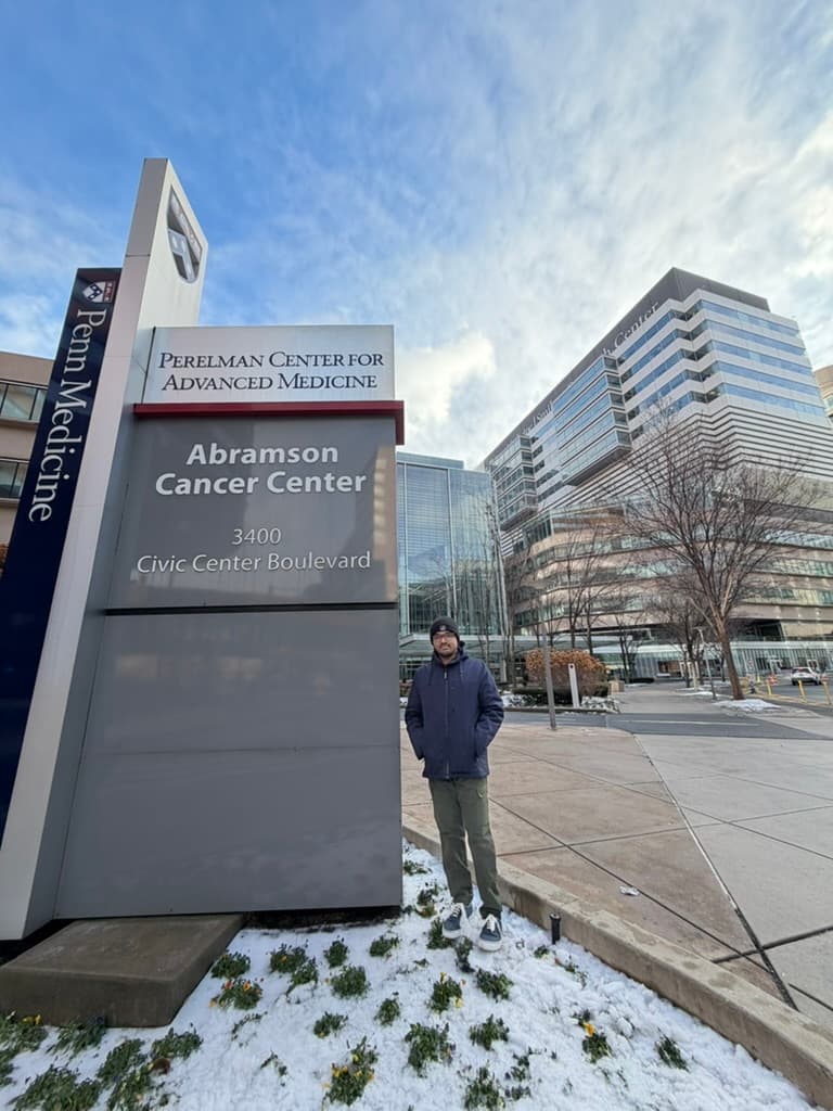
Began working on AI-assisted hemothorax detection in trauma CT, supported by NIH R01 funding through UPenn and the Reliance Foundation Undergraduate Scholars Program.
Lab pageMy research research contributes to AI4Healthcare, AI4Science, and trustworthy AI systems.
01 — University of Pennsylvania
Advisor: Prof. Walter Witschey
Ensembling 2D and 3D architectures for internal chest bleeding detection in trauma CT
Hemothorax, internal chest bleeding from trauma, requires rapid diagnosis, but manual CT analysis is slow and subject to human variability. I developed an AI-assisted detection system at Penn Medicine to help radiologists identify these critical bleeds more quickly and consistently. The system combines multiple deep learning architectures working together: 2D models analyze slice-by-slice patterns while 3D models capture full volumetric context. By merging their predictions, this ensemble approach outperformed both individual models and fine-tuned versions of existing medical imaging systems. The work shows that architectural diversity, not just more data, can drive better clinical AI performance. This reduces diagnostic delays in emergency settings and gives radiologists a reliable AI assistant for detecting these life-threatening bleeds.
Read full thesis02 — Carnegie Mellon University
Advisor: Prof. Min Xu
Discovering molecular structure in cryo-ET without a single human annotation
Cryo-electron tomography generates 3D images of molecular structures inside cells, but manually labeling these volumes is prohibitively slow, creating a bottleneck for biological discovery. I built and compared two unsupervised pipelines for grouping similar structures without human annotation. The first paired a 3D ResNet-18 pretrained on video recognition with t-SNE for dimensionality reduction and K-Means clustering, which requires a fixed cluster count set in advance. The second trained a 3D SimCLR encoder from scratch using contrastive self-supervised learning, paired with UMAP for manifold learning and HDBSCAN, a density-based method that determines its own cluster count and separates out noise. The self-supervised pipeline produced substantially better cluster coherence and cleaner separation between groups. Because the encoder, dimensionality reduction method, and clustering algorithm all changed together between the two pipelines, this compares the two pipelines as a whole rather than isolating self-supervision as the sole cause.
03 — MIT Media Lab
Advisor: Prof. Ramesh Raskar
Keeping SONAR, a peer-to-peer federated learning framework, reliable as it scaled
Contributed to SONAR (Self-Organizing Network of Aggregated Representations) at MIT Media Lab, a decentralized federated learning framework enabling peer-to-peer collaborative model training across distributed nodes without centralizing raw data. Refactored core algorithm modules and utility services to enforce consistent coding standards, introducing static analysis tooling via Pylint and Pyright with enforced score thresholds and pre-commit hooks to catch regressions at the source. Hardened the CI/CD pipeline by authoring GitHub Actions workflows covering automated linting, type checking, and functional end-to-end training tests over the gRPC communication backend, ensuring new contributions remained reliable as the codebase scaled across both MPI and gRPC deployment environments.
Independent research · ongoing
In progress
Making VLM radiology reports auditable rather than merely plausible
A vision-language model will write a fluent radiology report and localise findings it never actually saw. The fluency is the danger: a plausible report is indistinguishable from a correct one until someone checks it. AuraRad treats a generated report as a hypothesis and builds the checking in.
The LLM orchestrates and writes; deterministic tools compute.
A CT study is unzipped and rendered client-side through Cornerstone3D with HU-accurate windowing, measurements and 3-plane MPR. Nothing is persisted, no PHI at rest. Most VLM endpoints accept a single image, so the study is stitched into one 2048×1024 frame: a labelled 16-slice collage beside the active high-resolution slice, with the prompt explaining the layout so slice references map back to real indices.
1 / 4 · Study
A research prototype, not a validated medical device, with no HIPAA controls. The disease classifier is a keyword stub awaiting a real model; most current VLMs will not return CT bounding boxes, so approximate markers dominate and true localisation needs a detection model.
Completed with their graded assignments and quizzes. Every entry links to its certificate.
Click on the logos to see pictures.
Location
University of Pennsylvania
Pennsylvania 19104, USA
Get in touch
Send an email